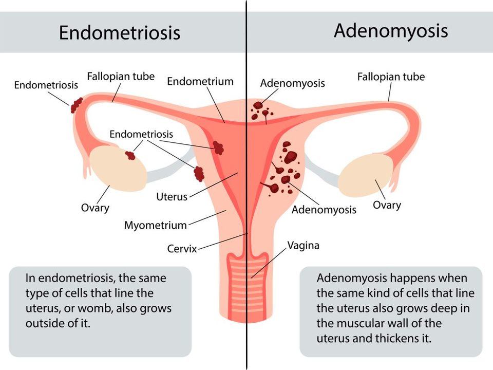
It usually starts with an exam. An enlarged, globular,
tender uterus is often the first clue — and the reason imaging gets ordered at
all.
Then a transvaginal ultrasound, which is almost always the
first test you’ll be given: painless, no radiation, widely available. Radiologists
work from a checklist of features, and the most telling one is a pattern called
echogenic subendometrial lines and buds. No single sign settles it.
The problem is what ultrasound misses. Pooled across
studies, transvaginal ultrasound finds adenomyosis in roughly seven out of ten
women who actually have it — so somewhere around one in four is
missed. That isn’t a rare failure. It’s a known limit of the
test.⁶
If you were told your ultrasound was normal and your symptoms
haven’t changed, that is a reason to ask for an MRI — not a reason
to accept that nothing is wrong.
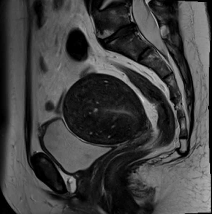
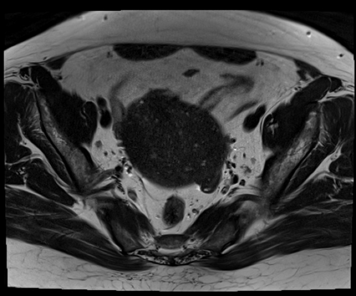
MRI is the more reliable test, and it’s what we use to
plan. It measures the junctional zone — the border between lining and
muscle, and often the earliest sign there is. It separates adenomyosis from fibroids,
which matters because nearly half of women have both. And it gives us the map we need
before an embolization. Short of taking tissue, it is the most sensitive test
available.⁶
Tissue is the one thing more definitive — and it’s also the
reason adenomyosis went unrecognized for so long. For decades the diagnosis was made by
a pathologist examining a uterus after it had been removed. Getting the
answer should not require losing the organ. That’s the whole case for
imaging it properly.