Understanding embolization

What embolization is, how it works, and what actually goes in.

Almost everything we do here is a form of embolization — for fibroids, arthritic knees, hemorrhoids, varicoceles, pelvic congestion. This page explains what the word means, why blocking a blood vessel on purpose can shrink a growth or quiet a painful joint, where the idea came from, and exactly what we put into the vessel to do it. If you’re reading about one of our procedures and wondering “but what actually goes in?” — this is the page.

The word, and the idea

An embolus is a plug. Embolization is placing one exactly where it helps.

Every part of your body has a blood supply. When something goes wrong — a fibroid growing, a joint lining inflamed, a hemorrhoid swollen, a vein letting blood flow backwards — that blood supply is very often what keeps the problem going. Extra blood feeds growth. Extra blood carries inflammation, and the extra nerve endings that come with it. Pooled blood stretches a vein wider every year.

Embolization turns that supply down, or off — from the inside.

Here’s how. Through a pinhole in the skin — usually at the wrist or the groin — an interventional radiologist threads a tube thinner than a phone-charger cable into the blood vessel. Live X-ray shows exactly where it is. A little contrast dye lights up the vessels, so we can see the abnormal ones: the tangle feeding a fibroid, the blush of new vessels around an arthritic joint.

Then we release the embolic — the plug — into that vessel, and only that vessel. Blood flow to the problem drops. Blood flow to everything around it stays.

The tube comes out. Pressure or a small closure device seals the pinhole. No incision, no stitches. Most patients go home the same day, and the pinhole is healed in days.

Catheter, in the main artery Embolic released here Healthy branch — keeps flowing Healthy branch — keeps flowing The problem
Simplified. The catheter is advanced past the branches that feed healthy tissue before anything is released.

Where does the blood go? Your body has backup routes — doctors call them collateral vessels. When one small branch is blocked, the healthy tissue next to it simply draws from another. That’s why the supply to a fibroid can be cut off while the uterus keeps its own, and why the abnormal vessels in an inflamed knee lining can be closed while the knee itself keeps its full blood supply. The target is chosen so that only the problem loses its feed.

Notice what embolization is not. Nothing is cut out. Nothing is opened. The organ or joint stays where it is, with its own blood supply from other routes. The problem inside it is simply starved of the extra blood that was feeding it. Why we call it “non-surgical”

One technique, many targets

What we embolize, and why.

The target and the goal decide everything else — which vessel, which agent, and whether the block should be permanent or temporary. Here is the same idea applied to each thing we treat.

Arteries · permanent

Uterine fibroids and adenomyosis

Target: the uterine arteries, and the branches feeding the fibroid or the thickened muscle. Goal: cut off the growth’s supply so it shrinks and softens, and the heavy bleeding eases. The uterus keeps its own blood supply from other routes. Agent: permanent microspheres, sized to the vessels.

Arteries · temporary

Painful, inflamed joints — knee, shoulder, hip, heel

Target: the abnormal little arteries that grow into an inflamed joint lining or tendon and carry the pain signal with them. Goal: calm the inflammation and quiet those extra nerve endings — while the healthy arteries to the joint stay open. Agent: a temporary slurry that dissolves within the hour. Nothing permanent stays behind.

Arteries · permanent

Hemorrhoids

Target: the branches of the superior rectal artery that carry too much blood into the hemorrhoid cushions. Goal: turn the excess inflow down so the cushions shrink and stop bleeding — done from upstream, with nothing at the anus at all. Agent: tiny coils, particles, or both.

Veins · permanent

Varicocele

Target: the testicular vein, which has faulty valves and is letting blood flow backwards and pool. Goal: close that vein for good so blood reroutes through the healthy ones and the pooling stops. Agent: coils to close the trunk, plus a sclerosant — a medicine that seals the vein wall.

Veins · permanent

Pelvic congestion

Target: the ovarian and pelvic veins that reflux and pool, the way varicose veins do in the leg. Goal: the same — close the leaking veins so blood finds the healthy route. Agent: a medicated foam that seals the tangle of smaller veins, and coils that close the main trunk.

In the hospital

Bleeding, tumors, malformations

Interventional radiologists also use embolization every day in hospitals — to stop internal bleeding after an injury or an operation, to starve liver tumors (often carrying chemotherapy in with the embolic), and to close aneurysms and tangled malformations of blood vessels. Same tools, same idea. Our practice focuses on the planned, outpatient procedures above.

Where it came from

Not new. Older than the MRI scanner — older than the CT scanner, too.

Patients sometimes ask whether embolization is experimental. The idea is nearly a century old, and it has been a routine tool in hospitals since the 1970s. What’s newer is where we’ve learned to apply it.

  1. 1930

    The idea appears

    An American surgeon is usually credited with the first deliberate embolization: treating an abnormal connection between an artery and a vein behind the eye by introducing a piece of muscle into the artery. Crude — but the principle was born. Block a vessel on purpose to fix a problem.

  2. 1953

    The pinhole

    Swedish radiologist Sven-Ivar Seldinger works out how to get a catheter into a blood vessel through a simple needle puncture, with no incision. Every embolization since starts exactly this way — it’s still called the Seldinger technique.

  3. 1960

    Delivered through the bloodstream

    Alfred Luessenhop treats a malformation of brain vessels by floating small spheres into the artery feeding it — the first embolization carried to its target by the blood itself.

  4. 1964

    Interventional radiology is born

    Charles Dotter opens a blocked leg artery from the inside — the first angioplasty — and proves that catheters can treat, not just take pictures. The specialty that does everything on this website grows from here.

  5. 1972

    Stopping bleeding without surgery

    Josef Rösch, working with Dotter, controls life-threatening stomach bleeding by embolizing the artery. Embolization becomes an emergency tool in hospitals worldwide — and still is, every night.

  6. 1975

    Coils and particles

    Cesare Gianturco introduces the metal coil, and the same decade brings gelatin sponge and PVA particles into use as embolics. The modern toolkit takes shape.

  7. 1978

    Veins, too

    Doctors in Portugal embolize the testicular vein to treat varicocele — the direct ancestor of the varicocele embolization we do today.

  8. 1990s

    Platinum coils, and fibroids

    Soft platinum microcoils arrive (1991). In 1995, Jacques Ravina in Paris reports embolizing the uterine arteries to treat fibroids — and women begin avoiding hysterectomy. Calibrated microspheres, made in exact sizes, follow shortly after.

  9. 2000s

    Fibroid embolization becomes standard

    Microspheres receive FDA clearance, including specifically for fibroids (2002). Liquid embolics for complex malformations follow. Uterine fibroid embolization becomes an established alternative to hysterectomy.

  10. 2013–2015

    Joints and hemorrhoids

    Japanese interventional radiologist Yuji Okuno reports embolizing the abnormal vessels around inflamed tendons (2013) and arthritic knees (2015) with a temporary agent — the start of knee, shoulder, hip, and plantar fascia embolization. In France, Vincent Vidal describes hemorrhoid artery embolization (2014).

  11. Today

    An outpatient specialty of its own

    Embolization is performed every day worldwide, and increasingly in dedicated outpatient suites like ours rather than hospitals. The tools are better made and better sized than ever. The idea hasn’t changed since 1930.

What actually goes in

The plug comes in many forms. Here is each one, plainly.

“Embolic” just means the material we use to block the vessel. Different jobs need different plugs. Some need to stay forever; some should dissolve. Some need to fill one large vein; some need to reach the tiniest branches. Here is what’s in the toolkit — the ones we use here, and a few you’ll read about elsewhere — and where you’d meet each one.

Particles — permanent

Microspheres and PVA particles Embosphere · Embozene · PVA

What they are. Tiny particles of a soft, inert material, made in graded sizes. Microspheres are perfectly round beads — a gelatin-based polymer in Embosphere, a hydrogel in Embozene. PVA particles are irregular flakes of polyvinyl alcohol, the original embolic particle from the 1970s, and still in regular use. The sizes are measured in microns, thousandths of a millimeter. To give you a sense: the largest we use are about the size of a grain of table salt; the smallest, a fraction of that.

How they work. Delivered through the catheter suspended in contrast dye, they float downstream until each one lodges in a vessel of its own size, and stays. Because the size is exact, we choose how deep into the vessel tree they travel — smaller spheres go further into the fine branches, larger ones stop sooner.

Where you’d meet them here. We use all three regularly. Fibroid embolization and UAE for adenomyosis use microspheres — the same spheres, just sized differently for the finer vessels of adenomyosis — and particles are one of the options for hemorrhoid embolization. Do they stay? Yes, permanently. They are made to be inert, and microspheres are FDA-cleared for fibroid embolization.1

Spheres and flakes behave a little differently: the exact sizing of spheres lets them travel a predictable distance into the vessel tree, while irregular PVA flakes tend to catch and pack a little sooner. Your physician picks the one that suits the vessel.

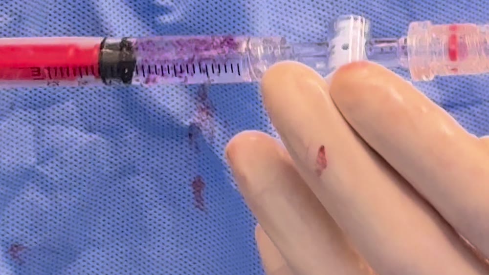
Watch the spheres go in. This is a syringe of Embozene microspheres, 900 microns each — just under a millimeter, among the largest sizes we use, which is why you can see them by eye — suspended in contrast dye and pushed through the stopcock into the microcatheter. The catheter carries them the rest of the way to the target vessel. Filmed in our suite; five seconds, looping, no sound.
Close-up of a syringe barrel holding hundreds of small purple microspheres suspended in clear contrast, next to the catheter hub
The same syringe, up close. The manufacturer tints the spheres so they can be seen and sized at a glance. Smaller sizes — used for finer vessels — look like a faint haze rather than individual dots.

Coils and plugs — permanent

Coils stainless steel, often fibered

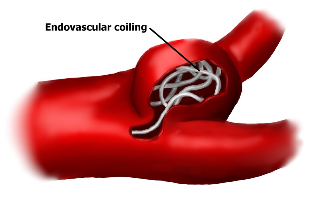
What they are. A very fine wire — the ones we use are stainless steel, often with tiny synthetic fibers along it to encourage clotting — that is pushed out of the catheter and curls into a coil a few millimeters across as it emerges. Several are packed in until the vessel is filled.

How they work. Blood clots around the coil, and the vessel closes. Over the following weeks the closure becomes permanent scar. Coils are the right tool for closing one larger vessel — a vein trunk, a main feeding artery — rather than a network of tiny branches, which particles handle better.

Where you’d meet them here. Varicocele embolization and pelvic congestion, where they close the faulty vein trunk; hemorrhoid artery embolization. Do they stay? Yes. You will not feel them. The stainless steel coils we use are not a concern for MRI scanning — we’ll give you a record of exactly what was placed so you can tell any imaging center.

A related device is the vascular plug: a single, self-expanding mesh plug that closes a larger vessel in one piece where many coils would otherwise be needed.

Medical illustration of fine metal coils packed inside a red blood vessel, labeled endovascular coiling
What coils look like in place. The fine wire curls as it leaves the catheter and packs the space until the vessel is filled; blood clots around it, and over the following weeks the closure becomes permanent. Medical illustration — not to scale.

Temporary agents — they dissolve

Imipenem/cilastatin slurry temporary crystals

What it is. Imipenem/cilastatin is a common antibiotic that comes as a powder. Mixed with X-ray contrast dye, it doesn’t fully dissolve — it forms a slurry of microscopic crystals, each smaller than a grain of sand.

How it works. Injected into the abnormal vessels around an inflamed joint or tendon, the crystals lodge in the tiny abnormal branches and shut them down — and then dissolve into the bloodstream, within about an hour. That is exactly what we want in a joint: enough of a block to close the abnormal vessels and calm the inflammation they feed, without leaving anything permanent in an artery that serves a joint you’ll use for the rest of your life.

Where you’d meet it here. Every musculoskeletal embolization we do — knee (GAE), shoulder, hip, and plantar fascia. Does it stay? No. And if you’re wondering how a temporary block gives lasting relief: in the study that compared these temporary crystals head-to-head against permanent beads for knee arthritis, relief at two years was just as good.2

Gelatin sponge Gelfoam

What it is. An absorbable gelatin sponge, first made in the 1940s to stop bleeding during surgery. For embolization it’s cut into small pieces, or whipped with contrast into a slurry, and delivered through the catheter.

How it works. It plugs the vessel for a matter of weeks, and then the body absorbs it. Its classic job is a temporary block that gives an injured vessel time to heal, or that backs up another agent.

Where you’d meet it here. Sometimes — we use it when a vessel needs a temporary block on its own or alongside coils. In hospitals it’s a mainstay for controlling bleeding. Does it stay? No — absorbed over a few weeks.

Sclerosants — for veins

Sodium tetradecyl sulfate Sotradecol

What it is. Not a plug at all — a medicine. A detergent-like liquid that irritates the inner lining of a vein so the vein seals shut and scars closed. It has been used on varicose veins since the 1940s. We usually whip it with a little air into a fine foam: foam pushes the blood aside so the medicine touches the vein wall, and it travels through a tangle of small veins that coils can’t reach.

How it’s used. Together with coils, in the veins. The coils close the main trunk; the foam handles the network of smaller veins around it. That combination is what our varicocele and pelvic congestion pages mean by “coils and a sclerosant” or “coils and a medicated foam.”

Where you’d meet it here. Varicocele embolization and pelvic congestion. Does it stay? No — it does its work on the vein wall and is absorbed. The closed vein itself shrinks to a cord over the following weeks.

Liquids — oils and glues (not typically used here)

Ethiodized oil Lipiodol

What it is. Poppy-seed oil bonded with iodine, so it shows up brightly on X-ray. First made in 1901 — the oldest thing in the toolkit.

How it’s used. On its own it slows blood flow in very fine vessels and lingers there. Its two big jobs: carrying chemotherapy into liver tumors (chemoembolization), and being mixed with glue so the glue can be seen on X-ray and its setting time controlled. A hospital-side agent — we don’t typically use it here.

Glue and liquid embolics n-BCA glue · Onyx

What they are. Medical “superglue” (n-butyl cyanoacrylate) hardens within seconds on contact with blood; mixed with Lipiodol, it can be seen and its set time tuned. Onyx is a liquid that solidifies from the outside in as it meets blood, spreading like slow lava into every branch of a vessel tangle.

How they’re used. To seal a bleeding vessel, a malformation, or an abnormal connection between an artery and a vein — places where particles or coils can’t get a complete seal. Both are permanent. They belong to the hospital side of the field — we don’t typically use them here, and you’re unlikely to meet them in a planned outpatient procedure.

Which agent, for which procedure, here

ProcedureWhat we typically useStays, or dissolves?
Uterine fibroid embolization · UAE for adenomyosisMicrospheresStay — permanent, inert
Knee (GAE) · shoulder · hip · plantar fasciaImipenem/cilastatin slurryDissolves within the hour
Hemorrhoid artery embolizationCoils, particles, or bothStay
Varicocele embolizationCoils + SotradecolCoils stay; the sclerosant is absorbed
Pelvic congestionCoils + sclerosant foamCoils stay; the foam is absorbed

This is what we typically use. Your physician chooses the agent and the size for your anatomy on the day, based on what the live X-ray shows — and will tell you exactly what was placed.

The honest part: the one risk every embolization shares. Whatever the agent, the main thing that can go wrong is material going somewhere it wasn’t meant to — doctors call it non-target embolization. That is why the whole procedure is done under live X-ray, why the catheter is advanced past the branches feeding healthy tissue before anything is released, and why the size and type of agent is matched to the vessel. Separately, many patients feel some aching, tiredness, and sometimes a low-grade fever for a few days afterward — that is the treated tissue reacting, and it is expected. Both get discussed plainly at your consultation, along with the risks specific to your procedure.

Sources. 1. U.S. FDA 510(k) K021397 — Embosphere Microspheres for uterine fibroid embolization, 2002. 2. Bhatia S, et al. Two-year outcomes comparing Embosphere microspheres versus imipenem/cilastatin for genicular artery embolization in patients with moderate to severe knee osteoarthritis. The Knee, 2023. Historical dates: Brooks (1930), Seldinger (1953), Luessenhop & Spence (1960), Dotter (1964), Rösch et al. (1972), Gianturco et al. (1975), Lima et al. (1978), Guglielmi et al. (1991), Ravina et al., Lancet (1995), Okuno et al. (2013, 2015), Vidal et al. (2014). Individual results vary. Nothing on this page is a promise about your outcome.

Straight answers

Questions patients actually ask about embolization.

“Does the tissue you block just die?”

Not the way it sounds. A fibroid loses its supply and shrinks and softens — that is the point, and the uterus around it keeps its own blood flow. Hemorrhoid cushions shrink but stay. In a joint we use a temporary agent, so the abnormal vessels close, the inflamed lining calms down, and the joint keeps its full supply. Healthy tissue is always fed by more than one route.

“Where does the blood go?”

Around. Your body has backup routes into every organ — collateral vessels. Block one small branch and the tissue next to it draws from another. The catheter is placed past those branches, so only the target loses its feed.

“Will I feel the coils or the beads?”

No. Coils are a few millimeters across and sit inside a vein or artery, well away from any nerve you can feel with. Microspheres are smaller than a grain of sand. Neither is something you can sense once you’ve healed.

“Can I still have an MRI?”

Yes. Microspheres, particles, and dissolved agents leave nothing that matters to a scanner. The stainless steel coils we use are not a concern for MRI either — we’ll give you a record of exactly what was placed, and you simply tell the imaging center.

“Is this experimental?”

No — and we’d rather be exact than vague. Embolization has been a routine hospital tool since the 1970s. Fibroid embolization has been done for thirty years. Varicocele embolization, longer. Joint embolization is the newest use, described in 2015 and now done in centers around the world — and each of our procedure pages tells you plainly how much evidence sits behind it.

“Am I asleep for it?”

No general anesthesia. Most procedures use light IV sedation with a dedicated sedation nurse — you’re comfortable, breathing on your own, and most patients say it feels like a strong margarita and wears off fast. Some vein procedures use a slightly deeper “twilight” sedation. Either way, you go home the same day.

“How much radiation is involved?”

Live X-ray is used in short pulses only while the catheter is being placed and the agent released — the same kind of imaging used for heart catheterizations every day. We keep the dose as low as we can and shield what isn’t being treated. Your physician can talk through the specifics for your procedure.

“Does anything stay in my body?”

It depends on the agent — and now you know which is which. Microspheres, PVA particles, and coils stay, permanently and inertly. The slurry we use in joints dissolves within the hour. Gelatin sponge and sclerosant foam are absorbed. The table above shows what we typically use for each procedure.

Free patient guide

Not ready to call? Take the guide instead.

Our free patient guides explain your condition, every treatment option — surgical and non-surgical — and the questions worth asking any doctor. Written by our physicians. No appointment required, and no follow-up phone call unless you ask for one.

We’ll email your guide plus occasional patient education. Unsubscribe anytime. Privacy policy.

Have a question this page didn’t answer? Ask the people who do this every day.

Call us directly, or schedule a consultation on our calendar below. You’ll sit down with a provider who will look at your imaging with you, tell you which procedure fits your case, and give you a plain answer about whether we can help. We do a complimentary benefits check when you come in — no need to call about your coverage beforehand.

210-405-1335

Consultation hours

  • Monday–Thursday8:00–5:00
  • Morning · last consult11:30 AM
  • Afternoon · last consult4:30 PM
  • FridayProcedures only

Closed on major holidays.

3212 Napier Park, San Antonio, TX 78231

Get directions

Stand-in image (Map © OpenStreetMap) — the live site embeds the interactive Google map here

Schedule a consultation

No referral needed for your initial consultation.

Rather book by phone? Call (210) 405-1335.