For San Antonio adults with one-sided low back pain that no scan has explained

“Your MRI looks fine.” You don’t feel fine. It may not be your spine.

The sacroiliac (SI) joints — where your spine meets your pelvis — are one of the most common and most-missed causes of long-running low back pain. They sit right where the spine gets blamed, and they don’t show up well on scans. At ARVC we don’t guess. An image-guided numbing injection tells us whether the SI joint is your pain source. If it is, we treat it — precision injections and radiofrequency ablation, in our office. If it isn’t, you leave knowing that too, and where to go next.

Complimentary benefits check before any procedure No referral needed for your initial consultation

A gray-haired man outdoors presses a hand to one side of his low back, just below the belt line
Below the belt line, to one side Classic SI joint pain sits low and off-center, and often runs into the buttock or the back of the thigh — exactly where the spine gets blamed for it.

Does this sound familiar?

It’s low — below the belt line — and it favors one side. Getting out of the car is the worst moment of the day. Rolling over in bed wakes you. Standing up from a chair takes a beat before you can walk straight. Long sitting, long standing, stairs, the wrong step off a curb — all of it lands in the same spot, and you can put a finger right on it.

You’ve done the work. The MRI of your back came back “unremarkable,” or showed a little wear that didn’t explain the pain. Physical therapy helped some. Maybe you had injections in your spine, or even a back procedure — and the pain is still there, in the same place. Somebody said “sciatica.” Somebody said “it’s your hip.” Nobody said “we’ve found it.”

And so you’ve started organizing your life around it: the pillow between your knees, the way you swing both legs out of the car together, the long drives you don’t take anymore.

Here’s what may have happened: the right joint never got tested. The SI joint can’t be diagnosed from a picture. It’s confirmed by numbing it — and that’s exactly what we do.

What the SI joint is

The joint between your spine and your pelvis. Strong, barely moving — and easy to miss.

You have two sacroiliac joints, one on each side, where the sacrum — the wedge of bone at the base of your spine — meets the two big hip bones of the pelvis. They’re wrapped in some of the strongest ligaments in the body and they move only a few millimeters. Their job is to hand the weight of your upper body down to your legs, every step, every day.

Like any joint, they can wear, strain, or get injured — and when one is irritated and inflamed, doctors call it sacroiliitis. The usual causes: years of load, a fall onto the tailbone, a lifting injury, pregnancy, a leg that’s a little shorter than the other, or the extra work an SI joint takes on after a spinal fusion above it. When one starts to hurt, the pain sits below the belt line, usually to one side, and often spreads into the buttock, the groin, or the back of the thigh — which is exactly why it gets mistaken for a disc, a pinched nerve, or the hip.

Here’s the part that keeps people undiagnosed. Studies put the SI joint behind somewhere between 15 and 30 percent of chronic low back pain¹ — and yet it’s a diagnosis of exclusion. It doesn’t reliably show on MRI. A lumbar spine scan is aimed at the spine. And a worn SI joint can look ordinary on a picture while a normal-looking one hurts. One of the most familiar patients in any SI joint clinic is the person whose spine has already been treated — sometimes very well — and who still hurts low and to one side.

BELT LINE L5 Sacrum Hip bone (ilium) Hip bone (ilium) SI joint SI joint where the pain usually sits
Where it is — and where it hurts Seen from behind: the sacrum sits between the two hip bones, and the SI joints are the seams on either side. The pain lives low, off-center, below the belt line.

Here’s how we find out

You can’t diagnose the SI joint from a picture. You numb it — and see what happens.

First, the exam. There’s a set of hands-on tests — pressing on the joint, loading it in certain positions — and when three or more of them reproduce your pain, the SI joint moves to the top of the list.¹ We’ll look at whatever imaging you have, and order more only if we need to rule something else out.

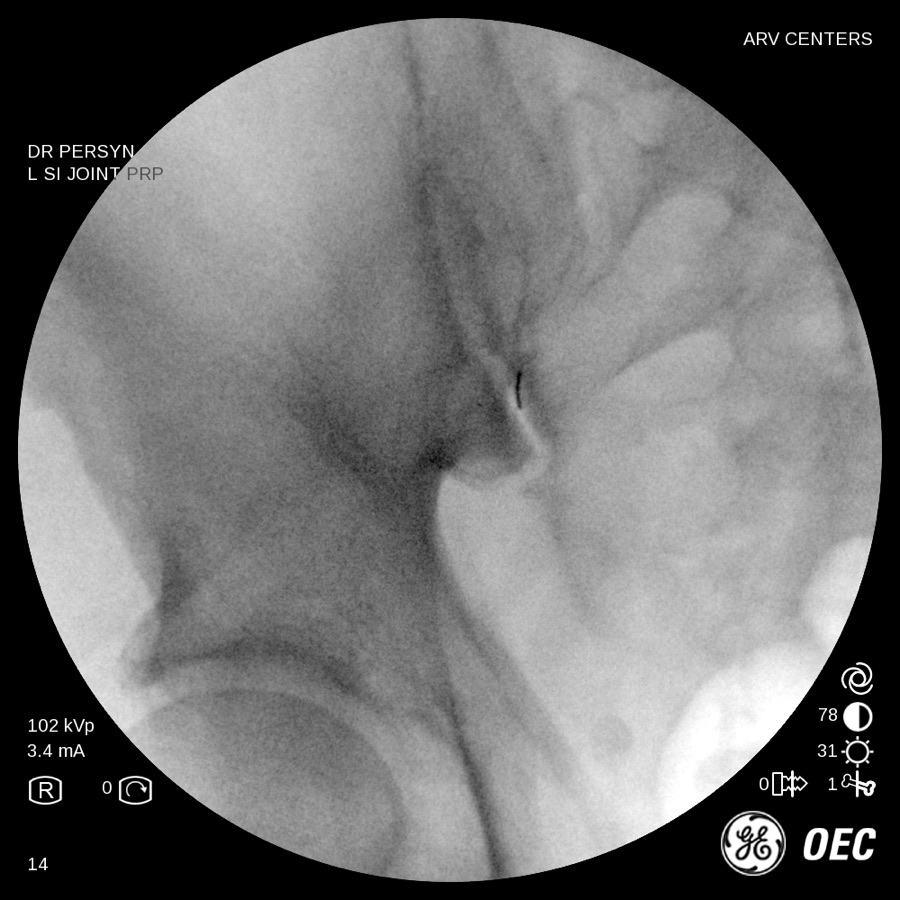
Then, the test that actually answers the question: a diagnostic block. Under live X-ray guidance, a thin needle is placed into the joint. A drop of contrast dye proves it’s in the right place — and that matters, because this is a relatively small joint buried under a lot of tissue, the thick gluteal muscle of the buttock. There is no reliable way to reach it by feel, and “close” isn’t good enough. Then we put in local anesthetic, usually with a dose of anti-inflammatory steroid. Now the important part happens on your end: over the next hours you pay attention. Did the pain drop most of the way — the movements that always hurt, suddenly not? If it did, the SI joint is your pain source. If it didn’t, it almost certainly isn’t — and you’ve just saved yourself months of treating the wrong thing.

When the answer isn’t clear-cut, we repeat it. Two blocks that agree are the most reliable test there is; even a single convincing block can fool you now and then, which is why we’d rather be sure before we go further.¹

So yes — your first procedure with us may be a diagnosis rather than a treatment. That’s by design. It’s a defined next step instead of a promise, and it’s the transparent way to treat a joint that scans can’t see.

Fluoroscopy arthrogram of an SI joint injection at ARV Centers, with contrast dye outlining the joint space
Image-guided means image-guided An SI joint arthrogram from an injection Dr. Persyn performed here: the contrast dye outlines the joint space, confirming the needle is in the joint — not near it.

Your options, in order

Least invasive first. Surgery last.

Injections to diagnose and to relieve pain. Ablation to make the relief last longer. And when those options fail, a referral for surgery — a last resort, and one we’ll be transparent with you about.

Physical therapy, anti-inflammatories, a support belt

Where everyone starts, and worth doing well: strengthening the muscles that stabilize the pelvis, and a sacroiliac belt for the days it’s bad. For a strained joint that’s sometimes enough. But if it were going to be enough for you, it would have been by now.

Image-guided SI joint injection — our first rung

The diagnostic block above, and treatment in the same needle: local anesthetic plus a steroid that calms the inflamed joint. Placed under live X-ray, in our office, in minutes. Relief typically lasts weeks to months, not forever, and it can be repeated when appropriate. We’ll be blunt about one thing the research is blunt about: for lasting relief, injections rank behind ablation and fusion.³ So we use them for what they’re best at — confirming the joint and buying real, temporary relief — and not as the end of the road.

Radiofrequency ablation (RFA) — when the block confirms the joint

The pain from an SI joint travels along tiny sensory nerve branches that run across the back of the sacrum. Radiofrequency ablation uses a heated probe tip, placed under X-ray guidance, to quiet those branches so the pain signal stops getting through. It’s done in our office, with local anesthetic only. In the published studies, relief lasts months, and for many people up to about a year¹ ⁴ — the nerves slowly grow back, which is why the effect fades and why it can be repeated. It doesn’t touch the nerves that move your legs. It’s the rung for people whose block said “yes” and whose injections stopped holding.

SI joint fusion

Here’s the transparent part: a surgery that locks the joint with implants ranks at the top of the comparisons for lasting pain relief.³ (Several of those trials were funded by the companies that make the implants, which is worth knowing.) We don’t perform it. If your joint has been confirmed, and ablation doesn’t hold, we’ll say so and refer you to a spine surgeon who does — with your blocks and your history in hand, so nobody starts over. It’s the last rung, not the first stop.

What to know first

Where the evidence stands — plainly.

Here is what the research says, plainly. Injections into the SI joint work, but they don’t last as long as radiofrequency ablation, and neither lasts as long as fusion surgery. When researchers pooled the head-to-head trials in 2023, that was the order — and they added that the evidence still “cannot lead to a definite conclusion.”³ We’re telling you this because it explains exactly how we work: we start with the least invasive option and move up only as needed. Injections to diagnose and to relieve pain. Ablation to make the relief last longer. And when those options fail, a referral for surgery — as a last resort.

15–30%

The share of long-running low back pain that traces to the SI joint, according to the research. Common — and still one of the most-missed diagnoses in back pain, because it can’t be seen on a scan.¹

3 or more

Hands-on tests that reproduce your pain in the exam room. When three or more point to the SI joint, a diagnostic block is likely to confirm it — which is why the exam comes first and the needle second.¹

Up to 1 year

How long relief from radiofrequency ablation of the SI joint has been shown to last. Some patients get less; the nerves grow back; and the evidence past twelve months isn’t there, so we don’t claim it.¹ ⁴

We do a complimentary benefits check before any procedure. No surprises.

Sources: 1. Cohen SP, Chen Y, Neufeld NJ. Sacroiliac joint pain: a comprehensive review of epidemiology, diagnosis and treatment. Expert Rev Neurother 2013;13(1):99–116. 2. Cohen SP. Sacroiliac joint pain: a comprehensive review of anatomy, diagnosis, and treatment. Anesth Analg 2005;101(5):1440–53. 3. Liu Y, Suvithayasiri S, Kim JS. Comparative efficacy of clinical interventions for sacroiliac joint pain: systematic review and network meta-analysis. Neurospine 2023;20(3):997–1010 (9 randomized trials, 652 patients). 4. Systematic review and meta-analysis of the effectiveness of radiofrequency ablation of the sacroiliac joint. Curr Pain Headache Rep 2024. Full texts before any print or ad use.

What it’s like

In our office. Home the same day.

Both the injection and the ablation happen in our own procedure room, in our office — not a hospital. You lie face-down. The skin over the joint is numbed. Using live X-ray, the doctor guides a thin needle to the joint — or, for ablation, a slim probe to the nerve branches beside it — and confirms the position before anything else happens. The injection itself is measured in minutes. Ablation takes longer, because each nerve branch is treated in turn, but you’re still home the same day.

After an injection: take it easy for 24 to 48 hours — nothing strenuous — and pay attention to your pain for the rest of the day; that’s the data we need. Some people feel a brief flare before the steroid settles in over a few days. After ablation, expect the treated area to be sore for a few days to a couple of weeks while the nerves quiet down; the relief builds over the following weeks rather than the same afternoon.

Both are done with local anesthetic only — no IV, no sedation, for the injection and for the ablation alike. That means no fasting, no driver: you drive yourself home. And you’ll hear from us the next day either way.

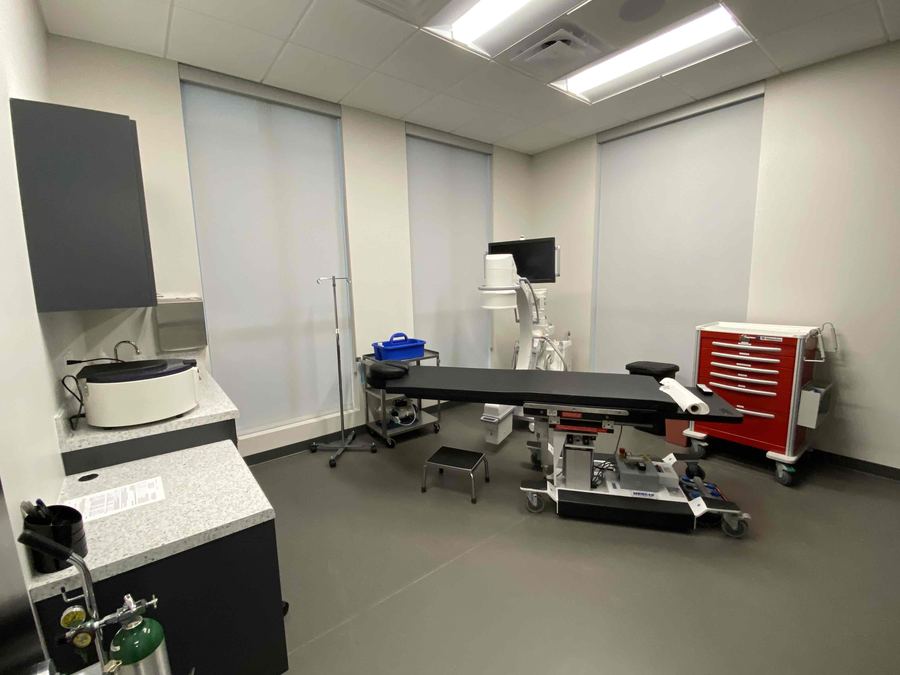
The procedure room at ARV Centers: a procedure table with a C-arm X-ray unit and monitor beside it, under natural light
Our procedure room — in the office The C-arm at the head of the table is the live X-ray that guides every SI joint needle. Not a hospital. Home the same day.

Candidacy

Who this is for — and who it isn’t.

You may be a candidate if:

  • Low back pain that sits below the belt line and favors one side has lasted three months or more
  • Your spine has been imaged, treated, or even operated on — and the pain is still there, in the same spot
  • Rolling over in bed, getting out of the car, or standing up from a chair are the worst moments
  • You’ve had a lumbar fusion, and new pain has shown up lower and to one side
  • It started after a fall, a lift, or a pregnancy — or crept up with years of wear — and nobody has actually tested the SI joint
  • SI joint injections helped once but keep wearing off, and you want to know what’s next

You’re probably not a candidate if:

  • The pain shoots below the knee with numbness or tingling — that’s usually a nerve in the spine, and we’ll say so
  • The diagnostic block doesn’t relieve your pain — if the block says no, we say no, and we tell you where we’d look next
  • You have warning signs — fever, unexplained weight loss, new bladder or bowel changes, pain after a serious injury — which need prompt evaluation elsewhere, not an injection

We turn away patients who aren’t candidates for what we do. Routinely.

Your care team

One team, under one roof.

Your exam, your diagnostic block, and your treatment all happen with our physical medicine team, in our office. Lisa D. Persyn, M.D., Board Certified Physical Medicine & Rehabilitation, performs your image-guided SI joint injections and your radiofrequency ablation — she has more than 100,000 joint injections behind her — and nurse practitioner Meredith Reynolds handles your exams and care through the visits. If your joint ever needs a surgeon, we’ll say so — and send your results with you.

Meet the whole team →

After your procedure

You’re not on your own.

We call you the next day — partly to check on you, and after a diagnostic block, partly because how the rest of that first day went is the answer we’re both waiting for.

Questions in between? You call, we answer. And if something ever feels urgent after hours, our phone line has an emergency option that rings the on-call doctor’s cell phone directly — any hour, any day.

Straight answers

Questions patients actually ask.

“My MRI was normal. How can anything be wrong?”

Because the MRI was probably of your lumbar spine, and the SI joint sits next door. Even when the SI joint is imaged, a worn joint can look ordinary, and a normal-looking joint can hurt. That is why the joint is diagnosed by numbing it, not by looking at it. Bring the scan anyway; it helps us rule other things out.

“I already had injections or a procedure on my back, and I still hurt in the same spot. Could it be the SI joint?”

It could, and you would be in familiar company. One of the most common patients in any SI joint clinic is the person whose spine has already been treated, sometimes very well, and who still hurts low and to one side. Sometimes the SI joint was the problem all along; sometimes it was the second problem, unmasked once the first was fixed. Either way, a diagnostic block will tell us.

“How soon will I know if the block worked?”

The same day. The numbing medicine takes effect within minutes and wears off over a few hours, and that window is the test: we ask you to do the things that always hurt — get in and out of the car, roll over, sit and stand — and pay attention. If the pain drops most of the way and then returns as the numbing fades, that is a clear answer. The steroid that goes in with it works more slowly, over days, and that is the part that gives longer relief. We call you the next day to hear how it went.

“Will one injection fix it?”

We won’t promise that, because the research doesn’t. Steroid injections into the SI joint give many people real relief for weeks to months, and they can be repeated when appropriate. But in head-to-head comparisons, injections rank behind radiofrequency ablation and fusion for how long the relief lasts. So we use them for what they are best at: confirming the joint and buying real, temporary relief. If the joint is confirmed and injections don’t hold, ablation is the next rung, not another round of the same shot.

“How long does radiofrequency ablation last? Does it hurt the nerve?”

In the published studies, relief from SI joint ablation lasts months, and for many patients up to about a year; nobody has good evidence past twelve months, so we don’t claim it. The nerves treated are tiny sensory branches that carry pain from the joint, not the nerves that move your legs. They slowly grow back, which is why the effect fades and why the procedure can be repeated.

“Does the injection hurt?”

Less than most people expect. The skin is numbed first, the needle is thin, and the live X-ray means it goes exactly where it should the first time. Most people describe pressure, then a few seconds of sting as the medication goes in. The whole thing is measured in minutes, not hours.

“What happens if the block doesn’t relieve my pain?”

Then we have learned something important: the SI joint is probably not your pain source, and you have not spent months treating the wrong thing. That is the point of testing before treating. We will tell you what we think is next — the hip, the spine, or something else — and where to go for it, even when the answer is not us.

“I had a spinal fusion. Is this related?”

It can be. When part of the lower spine is fused, the joints below it carry more of the load, and the SI joint is the next joint down. New pain that sits lower and more to one side than the pain your fusion fixed is worth testing. We will need your surgical history and, ideally, your imaging.

“Is this an epidural?”

No. An epidural puts medication in the space around the spinal nerves, and we don’t do those here — no epidurals, no spinal injections. An SI joint injection goes into the joint itself, well below and to the side of the spine, and it answers a different question: is this joint the source? If your pain turns out to be coming from the spine, we will say so and point you to the right place for it.

“Is it covered by insurance?”

Image-guided SI joint injections are an established, broadly covered procedure. Radiofrequency ablation is usually covered too, once diagnostic injections have confirmed the joint; many insurers in fact require those blocks first, which happens to match how we work anyway. As always: we do a complimentary benefits check before any procedure. No surprises.


Free patient guide

Not ready to call? Start with the SI joint guide.

Our physicians wrote a plain-English guide to low back pain that isn’t the spine: how to tell SI joint pain from a disc, a nerve, or the hip; how a diagnostic block works; and what each treatment — injections, radiofrequency ablation, fusion — can and can’t do. No appointment required, and no follow-up phone call unless you ask for one.

We’ll email your guide plus occasional patient education. Unsubscribe anytime. Privacy policy.

Bring your imaging and your history. You’ll leave knowing whether the SI joint is your pain source — and what to do about it.

Spine MRIs, X-rays, the notes from anything already tried — bring it all. You’ll leave with a plan to find out for certain, and a straight answer about whether we can help — including a plain “this isn’t us, and here’s who it is” if that’s the truth. Call us directly, or send this form and we’ll call you to schedule. We do a complimentary benefits check before any procedure. No surprises.
210-405-1335

Consultation hours

  • Monday–Thursday8:00–5:00
  • Morning · last consult11:30 AM
  • Afternoon · last consult4:30 PM
  • FridayProcedures only

Closed on major holidays.

3212 Napier Park, San Antonio, TX 78231

Get directions

Stand-in image (Map © OpenStreetMap) — the live site embeds the interactive Google map here

Schedule an SI joint consultation

No referral needed for your initial consultation.

Rather book by phone? Call (210) 405-1335.