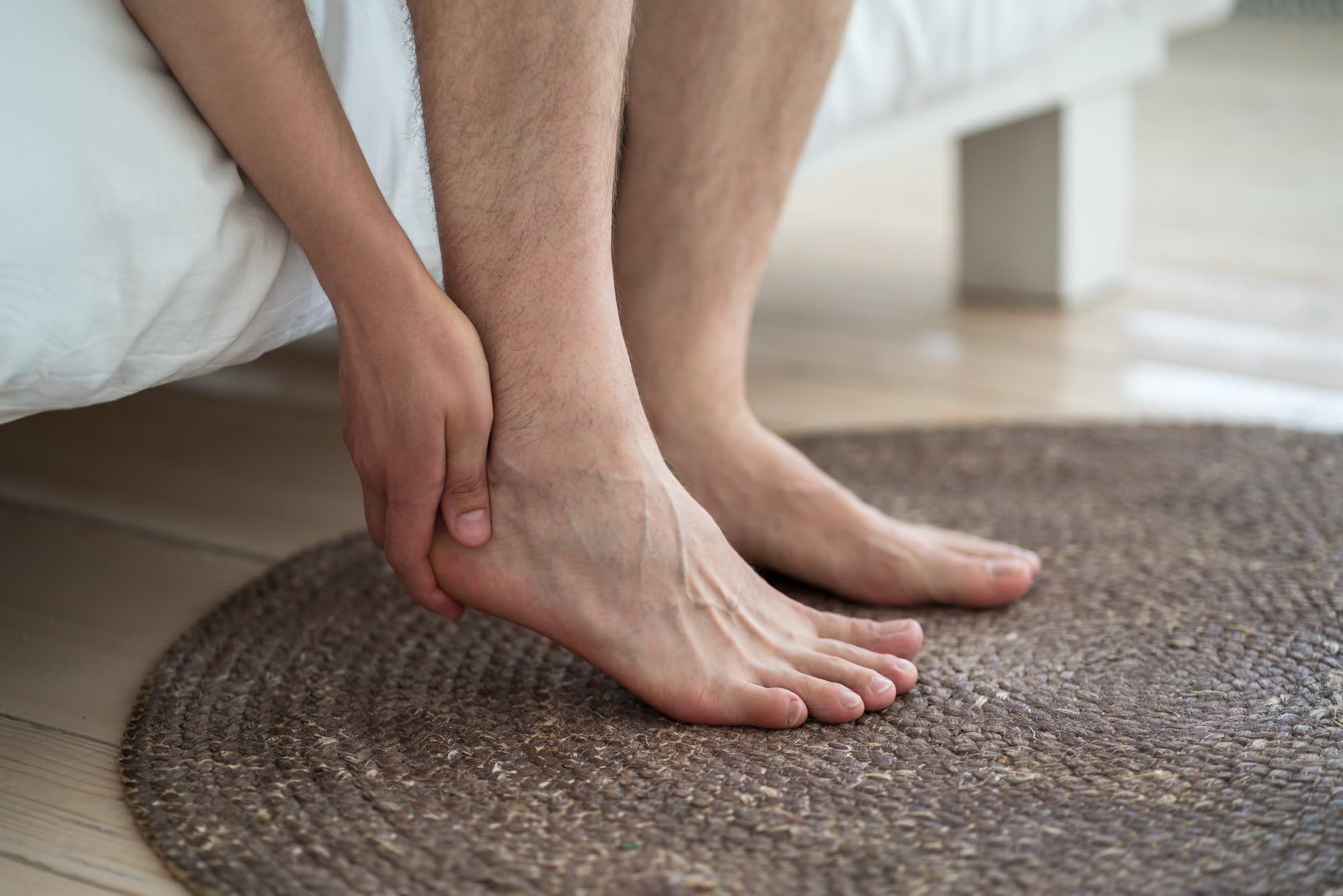
The first steps Out of bed, feet down, and the first thirty seconds are the worst part of the day.
Most heel pain gets better with the basics. If yours hasn’t — if you’ve
done the stretches, the therapy, the inserts, and the shots, and it still won’t let
go — you’ve earned a straight conversation about what’s left. Now, not
after another year. At ARVC that means an
exam that actually looks at your foot, an honest word on the evidence behind
every option, and two treatments we perform when they fit: image-guided PRP, and
plantar fascia embolization
— a same-day, no-incision procedure that quiets the abnormal blood vessels feeding
the inflamed tissue at your heel with a temporary agent that dissolves within the hour.
Nothing permanent stays behind.
Embolization typically covered by insurance — we check yours, free No referral or prior authorization needed for your initial consultation
Does this sound familiar?
You swing your legs out of bed, put your feet down, and the first thirty seconds are the worst part of your day. It eases as you get moving. Then you stand through a shift, or a grocery run, and it’s back.
You’ve done the calf stretches — everyone sent you the video. Then the inserts, then the custom ones. A night splint. A cortisone shot that helped for six weeks and then didn’t. Maybe shockwave, at $250 a session, out of pocket. Everyone had a different answer, and every appointment felt like waiting for it to go away on its own.
And now everything has a cost attached. The shortest route through the store. Deciding in advance where you can sit. The walks you’ve quietly stopped taking. And a foot is hard to explain — people think you’re being dramatic.
You’re not. This is a real condition with a published prognosis, and it deserves a real plan — with a date on it.
What’s actually wrong
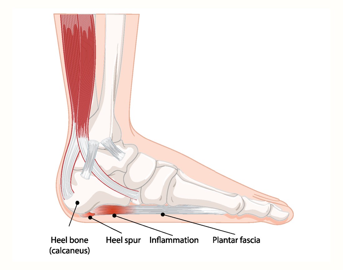
A thumbprint of tissue — and the spur isn’t the problem.
The plantar fascia is a thick band that runs along the bottom of your foot, from your heel
bone to your toes. It works like a bowstring: it holds up your arch, and every push-off
loads it. Plantar fasciitis is what happens where that band attaches to your heel. The
tissue there gets overloaded, breaks down, and hurts. It’s an area about the size of
a thumbprint, and it can take a life apart.
The name is slightly wrong, and that matters. “-itis” means inflammation, but
the current guideline is more careful: the problem runs along a spectrum with
both inflammatory and degenerative features.¹ Part is
inflammation. Part is tissue that has broken down and isn’t repairing. That’s
why ice, pills, and cortisone often help for a while and then stop — they treat one
half. And when tissue stays irritated long enough, the body grows abnormal new blood
vessels into it, and new pain nerves grow right along with them. That loop is what one of
our treatments targets.
Where it actually hurts
The band, heel to toes — and the thumbprint where it attaches. That’s the whole problem. The spur next to it is a bystander.
Sources: 1. Koc TA, et al. Heel pain — plantar fasciitis: revision 2023, clinical
practice guideline. J Orthop Sports Phys Ther 2023;53(12) — “may exist along a spectrum that includes both
inflammatory and degenerative characteristics.” 2. Hansen L, et al. Orthop J Sports Med 2018 — 174 patients
followed 5–15 years; heel spur at baseline had no effect on prognosis (p = .88).
How long this actually lasts
Two true numbers. Which one is yours depends on how long you’ve had it.
The first number is the one you’ll find everywhere, and it’s the reason the
conservative work comes first and gets a real chance. The second is the one nobody prints
— and it’s the reason we take a person who has done all of it and is still
stuck seriously, instead of telling them to wait another year.
More than 8 in 10
people with plantar fasciitis are completely better within 12 months.¹
46%
of people whose plantar fasciitis was bad enough to reach a specialist still had
symptoms an average of nearly ten years later. Not a forum post — a study of 174
patients.²
About 2 years
how long symptoms had lasted, on average, in the people from that study who
did recover. If you’re eighteen months in and improving slowly,
that’s not failure. That’s the shape of this thing.²
What we take from those
two numbers together. Early on, the odds are genuinely with you — do the
conservative work properly and give it a real, written-down trial. Done all of it and still
refractory? Then “wait longer” has stopped being a plan, and we don’t make
you serve out a year before we’ll talk. That’s when the rest of this page becomes
relevant.
Sources: 1. Viglione V, et al. EFORT Open Rev 2023 — systematic review
& meta-analysis, 42 double-blind trials, 1,724 placebo-arm patients: “over 80% of affected
patients gaining complete resolution within 12 months.” 2. Hansen L, et al. Orthop J Sports Med
2018 — 174 patients with ultrasound-confirmed plantar fasciitis followed 5–15 years (mean 9.7):
46% still symptomatic; risk of persistence 80% at 1 year, 50% at 5, 46% at 10; mean symptom duration in
those who recovered 725 days; heel spur and fascia thickness had no effect on prognosis.
First, the exam
Nobody should diagnose your heel without touching it.
Plantar fasciitis is diagnosed by history and examination — not by a scan. The
guideline lists what a proper exam looks for:¹ pain on the inside of the
heel, worst with the first steps after rest; tenderness when the examiner presses
the spot where the fascia attaches to the heel bone; a positive windlass
test — your toes are pulled back while you stand, which tightens the band and
reproduces the pain; negative tarsal tunnel tests, which is how a nerve
problem gets ruled out; and limited upward movement at the ankle.
Imaging supports the exam rather than replacing it. An X-ray, MRI, or ultrasound helps
confirm the picture and rule other things out — and if embolization is on the
table, ultrasound is how we see the thickened fascia and the abnormal vessels it
targets. The guideline is also specific about one more thing: reassess the diagnosis when treatment isn’t working.¹
Years of failed treatment is itself a reason to go back to the question. Things that get
mistaken for plantar fasciitis include a thinned heel fat pad, a stress fracture of the
heel bone, a pinched nerve under the heel or at the inner ankle, an inflammatory
arthritis, and a benign lump in the fascia. Most heel pain is plantar fasciitis. But
“is this definitely what I have?” is a fair question after a real run at
treatment, and a good clinician will welcome it.
Toes back, band tight
Pull the toes up and the fascia tightens — that’s the windlass test. It’s also the stretch that works.
Source: 1. Koc TA, et al. Heel pain — plantar fasciitis: revision 2023, clinical
practice guideline. J Orthop Sports Phys Ther 2023 — diagnostic criteria (medial heel pain worst with initial steps,
tenderness at the proximal insertion, positive windlass test, negative tarsal tunnel tests, limited ankle dorsiflexion) and
the recommendation to reassess when symptoms don’t fit or don’t respond.
Your options, in honest order
Every treatment — including the ones we don’t do.
The order below is roughly the order you should try things. Ours are near the end,
because that’s where they belong. Every option gets its weakness printed —
including all three of ours — and the same standard of proof applies to everyone.
1. The boring things — done properly, for twelve weeks
Most people say they’ve “tried stretching.” Almost nobody has done
the specific protocol, the way the trials did it, for as long as the trials ran. The
fascia-specific stretch — toes pulled back toward the shin, ten
holds, three times a day, the first round before you stand up in the morning
— beat the usual calf stretch in a trial of people already ten months in.¹
Add calf stretching, a loading program (slow heel raises every other
day, which sped recovery in a randomized trial),² a night splint if mornings are
your worst time, and an off-the-shelf insert — the trials found no
difference between $600 custom orthotics and store-bought ones at 6 weeks, 12 weeks, or
a year.³ Written down, with a follow-up booked at the end. If you haven’t done
this yet, we’ll tell you to — and show you how.
2. Hands-on therapy — where we send most people first
Manual therapy — mobilizing the foot and ankle, working the soft tissue of the
arch, heel, and calf — carries the guideline’s highest grade,
the same tier as stretching and higher than orthotics, laser, or dry needling.⁴ In a
randomized trial against custom orthotics, the manual-therapy group improved
most.⁵ It’s low-risk, usually covered, and if it’s going to work
you’ll know inside a month. We don’t perform this. In San
Antonio we usually refer people to Airrosti first, because in our
experience it’s often effective — and to be plain about it, we have no
business relationship with them. If you haven’t done this yet, it comes before
anything of ours.
3. Image-guided cortisone — one of the three we perform
We offer cortisone injections, and we’ll be straight with you about what they
do. Cortisone works quickly for some people and doesn’t hold: the
largest review — 47 randomized trials — found it no more effective than a
placebo injection for pain.⁶ What it can do is break a bad patch when you
can’t get through the day, and buy you the window to do the stretching and loading
that actually settles a heel. When we give one, it goes in under live
imaging — into the fascia at the heel, not near it. The rupture fear is
smaller than the forums suggest and attaches to repeated shots; one
image-guided injection is defensible. What we won’t do is give you a fourth one
and call it a treatment plan.
4. Shockwave and dry needling — we don’t offer these
Shockwave beats placebo, but the arithmetic matters: it drops pain
scores about 3 points out of 10, and in this condition a placebo alone produces 2 to
3.⁷ Real, modest, often not covered. Dry needling is graded above
orthotics and beat cortisone at long-term follow-up.⁶ We don’t perform
either one — and if one of them fits your heel, we’ll tell you so and point
you to someone who does.
5. PRP — one of the three we perform
Your own blood, spun down to concentrate the platelets that carry your body’s
repair signals, injected into the damaged tissue under ultrasound guidance. Across
24 randomized trials and 1,653 patients, PRP beat cortisone on pain at
three and six months and on function out to a year.⁸ It isn’t faster
— at one month the two were even — the advantage at twelve months was no
longer significant, and it’s sore for a few days afterward. $900 per
injection, cash. Insurance does not cover PRP. We’d rather print the price
than have you find it out in the room — and we’ll tell you plainly when
it’s worth considering and when it’s not.
Through a pinhole in an artery, an interventional radiologist finds the abnormal blood
vessels feeding the inflamed tissue at your heel and quiets them with a temporary slurry
of microcrystals that dissolves into the bloodstream within the hour — nothing
permanent stays behind. Normal blood supply to the foot is left alone. No incision, no
general anesthetic, home the same day, walking. Across four published series, pain
scores fell from 7–8 out of 10 to about 1–3, with no serious
complications — and in our own patients the results are just as encouraging.
It’s safe, well tolerated, and leaves nothing permanent behind. It fits people who have done the conservative work and are still stuck: for
them it can be the reliable, effective option they haven’t been offered — and
if that’s you, we don’t make you wait.
Two operations, sometimes combined: cutting part of the fascia to release its tension,
or lengthening the tight calf muscle instead. It works for most people who have it
— across 21 studies and 601 feet, pain fell from 7.8 to 1.8 out of 10.⁹ It
also isn’t free: 18.7% had a complication, and cutting the band
that holds up your arch is not reversible.⁹ Recovery is weeks to months. We
don’t perform this. If it’s your best option, we’ll say so and
tell you who to see.
Sources: 1. DiGiovanni BF, et al. J Bone Joint Surg Am 2006 —
plantar-fascia-specific stretching vs. Achilles stretching in chronic plantar fasciitis (>10 months). 2. Rathleff MS,
et al. Scand J Med Sci Sports 2015 — 48-patient RCT, high-load strength training vs. stretching, both
with inserts: faster improvement at 3 months, similar at 12. 3. Whittaker GA, et al. Br J Sports Med 2018 —
systematic review & meta-analysis, foot orthoses: no difference custom vs. prefabricated at 6 wk, 12 wk, 12 mo.
4. Koc TA, et al. Heel pain — plantar fasciitis: revision 2023, clinical practice guideline. J Orthop Sports
Phys Ther 2023 — manual therapy, stretching, night splints and taping graded A; orthotics alone graded
against for short-term relief. 5. Rodríguez-Sanz D, et al. Sports 2019 — RCT, manual therapy vs.
custom orthoses vs. both. 6. Whittaker GA, et al. BMC Musculoskelet Disord 2019 — 47 RCTs, 2,989
patients: corticosteroid injection “not more effective than placebo injection”; no ruptures in the
randomized data; dry needling superior at long-term follow-up. 7. Ramaccia et al. 2024, ESWT meta-analysis (~3-point
VAS drop) read against Viglione 2023 (placebo 2.1–2.8). 8. Zuo et al. Am J Phys Med Rehabil 2025 —
24 RCTs, 1,653 patients, PRP vs. corticosteroid. 9. Ward S, Shimozono Y, Kennedy JG. Foot Ankle Orthop
— systematic review of endoscopic treatment, 21 studies / 601 feet: pain 7.8→1.8, 18.7% complications.
Full options guide, including treatments not on this page, in the free patient guide below.
When injections aren’t enough
The next rung is plantar fascia embolization — what the studies say, and what we see.
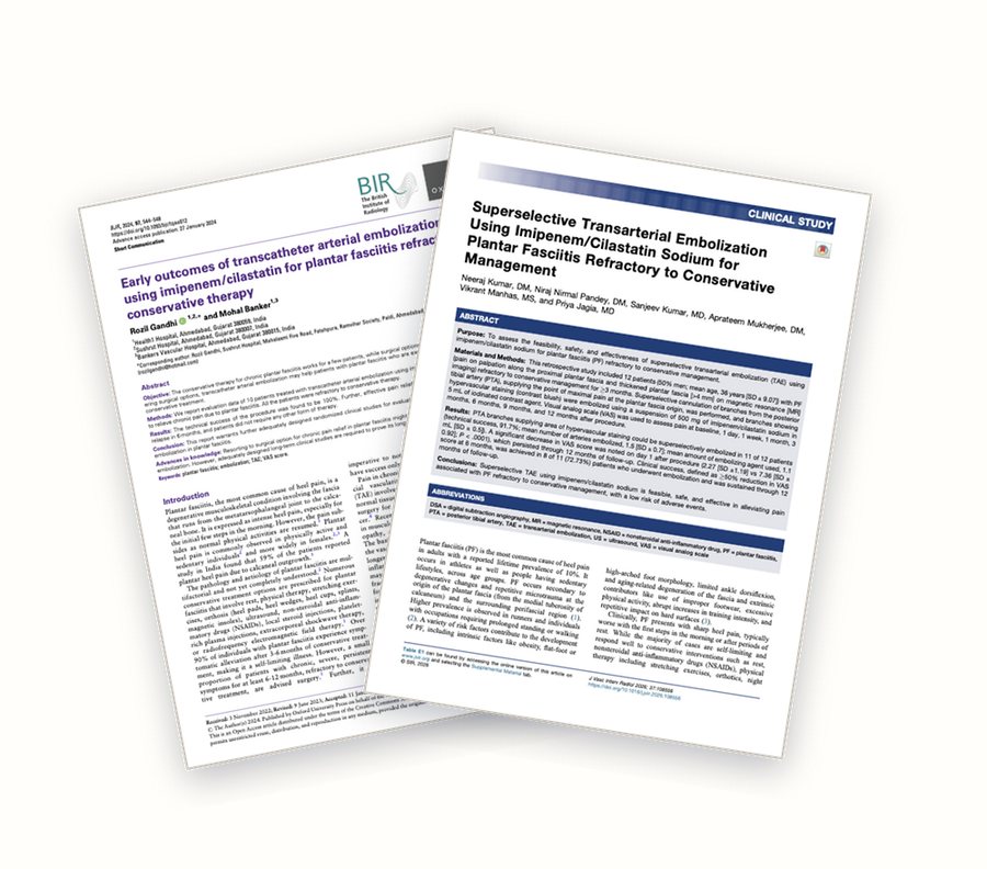
Two of the published heel series — both cited below. The record here is small and young: roughly 120 patients across four countries, and we say so plainly.
First, the good news, and it’s true for most people reading this: the great
majority of plantar fasciitis responds well to conservative care — the stretching, the
loading, the hands-on therapy, a supportive shoe. Done properly, those measures settle it for
most patients, and the likelihood of ever needing a procedure is small.
But some heels don’t respond. When the fascia has been inflamed long enough,
the body grows abnormal new blood vessels into the attachment at the heel, and pain nerves grow
in alongside them — and at that point the usual measures are treating the symptom while
the supply line stays open. Those patients need something more, aimed at the source of the
inflammation and the pain. That is exactly where plantar fascia embolization comes
in. It blocks those abnormal vessels; cut off the supply, and the inflammation settles
and the pain eases.
The published studies — roughly 120 patients across four countries, every one
of them with heel pain that had already failed conservative care — tell a consistent
story: pain scores fall from 7 or 8 out of 10 to about 1 to 3, function improves, the relief
holds, and there have been no serious complications. That matches what we see
in our own patients: in our experience the procedure is highly effective — most of
the patients we treat get real, meaningful relief. Here’s what the studies
show.
7.3 → 1.3
Average pain out of 10, before and six months after embolization, in the largest
prospective study — 32 patients whose heel pain had already failed conservative care.
Function scores more than halved. No complications.¹
7.9 → 0.7
Average pain at one year in the largest series — 66 patients, followed up to
four years, with the improvement holding at an average of 31 months. Some patients had
a second session.²
Zero
Serious complications reported across roughly 120 published patients — no tissue
damage, no ulcers, no fascia rupture. The side effects were bruising at the puncture
site.¹ ² ³ ⁴
Two smaller series used the same temporary agent we use. In India, 12 patients
saw pain fall from 7.4 to 2.6 out of 10 at a year, with 8 of 11 treated patients reaching at
least a 50% improvement and holding it, and no adverse events.³ In the UK, 10 patients
— all past three months of failed conservative care and averaging a year of symptoms
— went from 8.5 to about 1 by six months, with no complications.⁴ In every series the
shape is the same: relief that comes on over the first weeks, then holds.
Across every published series, the procedure has been safe and well
tolerated — and it leaves nothing permanent behind. For patients who have
genuinely exhausted their other options, that’s what makes it worth a conversation: a
safe, reliable, effective treatment that doesn’t involve cutting anything —
performed in our own office, by board-certified interventional radiologists, with a
temporary agent that dissolves within the hour.
We do a complimentary benefits check before any procedure. No
surprises.
Sources: 1. Tonkaz M, Bekci T. Eur J Radiol
2026;195:112573 — 32 patients, prospective single-center, superselective medial calcaneal artery,
imipenem/cilastatin; VAS 7.3→1.3 and FFI 55.5→20.4 at 6 months; fascia 5.2→3.7 mm; no major or minor
complications; minimal recurrence in 5/32 (physically demanding occupations), no repeat embolization; no control
group. 2. Sasaki T, Shibuya M, Miyazaki K, et al. Foot Ankle Surg 2025;31:105–110 — 66 patients,
ultrasound-guided non-selective embolization; NRS 7.9→0.7 at 12 months, AOFAS 65.8→92.8, maintained to mean
30.9 months; mean 2.3 sessions per patient; no major adverse events; uncontrolled. 3. Kumar N, Pandey NN, et al.
J Vasc Interv Radiol 2026;37:108556 — 12 patients, retrospective, superselective posterior tibial
branches, imipenem/cilastatin; technical success 11/12; VAS 7.36→2.27 at day 1, 2.64 at 12 months; ≥50% VAS
reduction at 6 months in 8/11, sustained to 12 months; no adverse events; single session; no control group.
4. Gandhi R, Banker M. Br J Radiol 2024;97(1155):544–548 — 10 patients / 13 procedures,
imipenem/cilastatin; VAS 8.5→1.9 at 1 month, 1.0 at 6 and 12 months; no complications; service evaluation.
A US study of the procedure is also under way (ClinicalTrials.gov NCT06805942).
What embolization is like
In our office. Home the same day — walking.
Before. A consultation, an exam, and an ultrasound of your heel.
We’re measuring the thickness of the fascia where it attaches and looking for the
abnormal vessels the procedure targets. If your scan doesn’t show that pattern,
we’ll say so — and point you to what will help instead.
The day. It’s done in our own IR suite, in our office — not
a hospital. Light IV sedation with a dedicated sedation nurse — most patients say
it feels like a strong margarita, and it wears off fast. Local anesthetic at a pinhole in the groin — no
incision, no stitches. A catheter narrower than a strand of spaghetti is guided to the
small arteries feeding your heel. Contrast dye shows the abnormal vessels as a blush the
normal side of your foot doesn’t have. The blocking agent is temporary: microcrystals
smaller than a grain of sand that quiet the abnormal vessels, then dissolve into the
bloodstream within 30 to 45 minutes — the same agent we use for knees and
shoulders, and the one used in the most detailed published heel series.² The normal
blood supply to the foot is left alone. About 45 minutes to an hour in the room, a short
observation, and someone drives you home the same day. Most people have mild bruising or
aching at the puncture site for a day or two.
The timeline nobody tells you. This is not a switch. Relief comes on over
weeks, as the inflammation settles and the tissue quiets down. In the published series,
most of the drop shows up in the first weeks and the rest builds through three and six
months.¹ If
you’re expecting to wake up fixed the next morning, you’ll be disappointed by
a procedure that may be working.
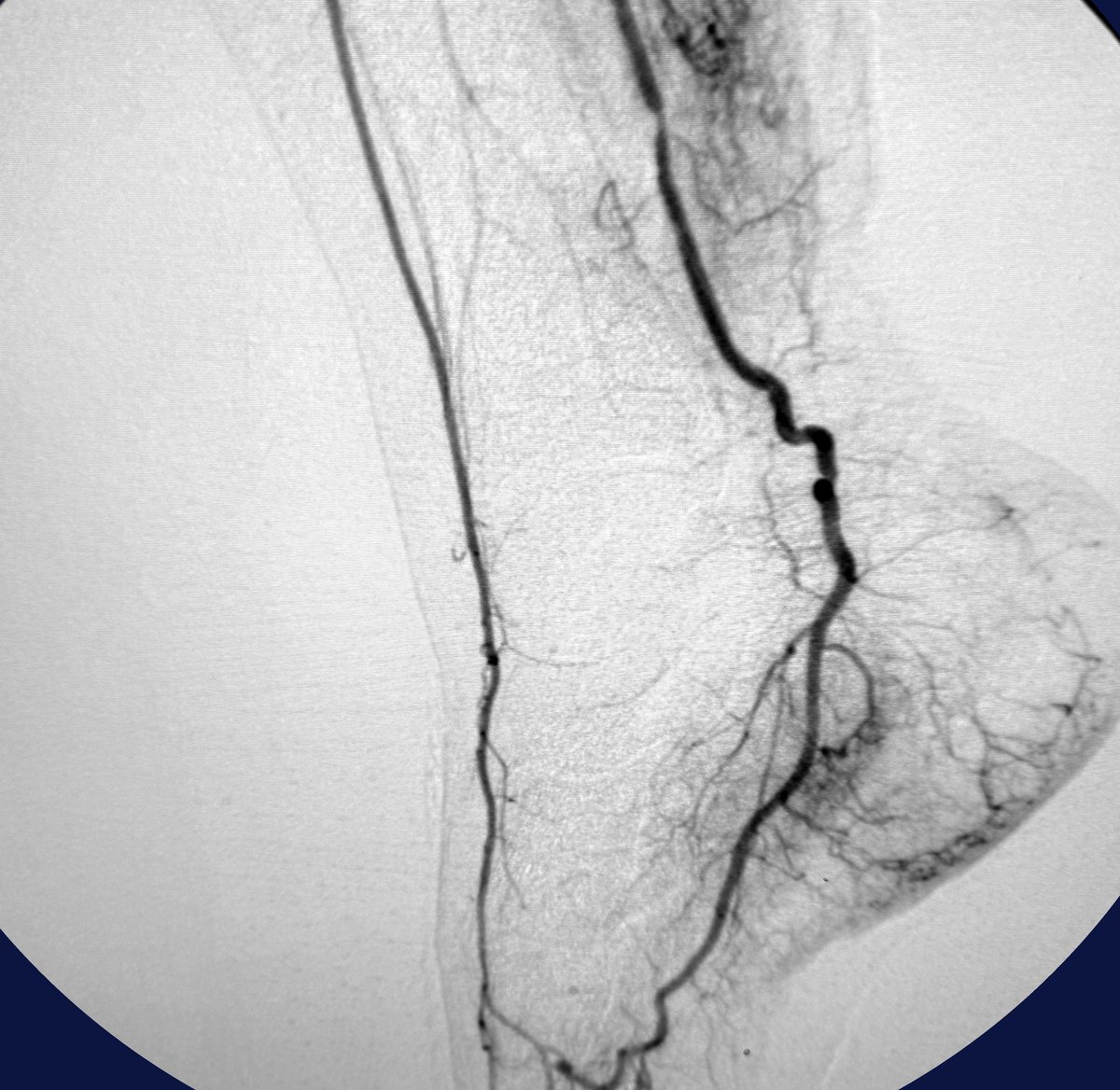
What PFE actually treats — one of our own cases
The artery runs down the inside of the ankle to the heel. The dark, tangled blush around the heel is the abnormal blood supply feeding the inflamed attachment. It’s what the catheter finds, and what the temporary agent quiets.
Sources: 1. Gandhi R, Banker M. Br J Radiol 2024 — VAS 8.5 at
baseline, 1.9 at 1 month, 1.2 at 3 months, 1.0 at 6 and 12 months; Kumar N, et al. J Vasc Interv Radiol 2026 —
VAS 7.4 at baseline, 2.3 at day 1, 3.5 at 1 month, 2.6 at 12 months. 2. Imipenem/cilastatin was the agent in Gandhi
2024, Kumar 2026 and Tonkaz 2026 — the three most detailed published heel series.
Candidacy
Who embolization is for — and who it isn’t.
You may be a candidate if:
You’ve given the conservative ladder a real run — stretching, loading, a splint, hands-on therapy — and you’re still refractory. There’s no waiting period after that: we’ll consider treatment right away
Ultrasound shows a thickened fascia at the heel and the abnormal vessels the procedure targets
Cortisone helped and stopped holding — or you’d rather not have another shot into tissue that’s already failed to respond
You want an effective option short of cutting the fascia — one that leaves nothing permanent behind
You’re probably not a candidate if:
You haven’t done the conservative work yet — more than 8 in 10 people get better without any of this, and we won’t skip it. You’ll leave with the twelve-week plan, and usually an Airrosti referral, instead
Your ultrasound doesn’t show the pattern the procedure treats — we’ll tell you, and talk through what will help instead
Your heel pain looks like something else — a stress fracture, a nerve, an inflammatory arthritis (we’ll look before we treat)
You have significant artery disease in the leg or foot
We turn away patients who aren’t candidates for what we do.
Routinely. And you still leave with a plan.
Your care team
One team, under one roof.
Your exam and, where they fit, your cortisone and PRP injections start with our
physical medicine team: Lisa D. Persyn, M.D., Board Certified Physical
Medicine & Rehabilitation, performs your image-guided injections — she has more
than 100,000 joint injections behind her — and nurse practitioner Meredith
Reynolds handles your exams and care through the visits. When embolization is the
right step, you’re handed to our Board Certified Vascular and Interventional
Radiologists. It isn’t a referral chain. It’s a hallway.
When you go home, you take the on-call doctor’s cell phone number with you.
Not an answering service — the doctor.
And you don’t have to wonder whether to bother us: we call you the next day
to check in. Every patient, every procedure.
Before you leave, you’ll get this card
Your procedure:
On-call doctor:
Cell phone: written by hand, before you walk out
Call me if anything worries you.
Straight answers
Questions patients actually ask.
“Is the heel spur causing this?”
Almost certainly not. In the study that followed 174 patients for up to 15 years,
having a spur made no difference to who got better. Plenty of people with a spur have
no pain at all. The spur is a bystander; the attachment is the problem.
“Will cortisone rupture my fascia?”
One injection: very unlikely. Across the randomized trials, no ruptures occurred. The
risk that shows up in clinic reviews attaches to repeated injections. It’s
a reason to be careful about the third and fourth shot — not to refuse the first.
“Do I need custom orthotics?”
The trials don’t show custom beating off-the-shelf at any point out to a year.
Try a store-bought insert first. If a $60 version helps, a $600 version is unlikely to
help more — and if you’ve already spent the money and felt let down, the
research agrees with you.
“Does embolization hurt? How long until I know if it worked?”
The pinhole is numbed; most people describe pressure rather than pain, and go home
the same day with mild bruising. Then: weeks, not days. In the published series,
improvement was measured at one month and kept improving through three and six.
We’ll give you a date to check in and a plan for what happens if it’s not
working.
“What if it doesn’t work?”
Then you’re where you were, minus a day — and surgery is still on the
table. Nothing has been cut, and nothing permanent was left behind. That’s the honest answer, and it’s a real part
of the decision.
“Why haven’t I heard of this?”
Because it’s a catheter procedure, and most foot specialists don’t do those.
Embolization is what interventional radiologists do — blocking abnormal blood vessels
that feed inflamed tissue — and it’s only in the last few years that it’s
been applied to the heel. The studies are recent, the results are consistent, and the
people who tend to hear about it are the ones who’ve run out of other things to
try. That’s exactly who it’s for.
“Can I have it in both feet?”
Bilateral cases are treated in the published series. We’d discuss timing with
you — and both feet get their own ultrasound, because both have to show the
target.
“Is it covered by insurance?”
Hands-on therapy and cortisone usually are, and embolization is typically covered
by insurance. PRP isn’t — $900 per injection, cash, at our published
price. Either way we don’t guess: we do a complimentary benefits check
before any procedure, usually the same day as your consultation, so you know
your number before you decide anything. If we don’t think a treatment is worth
your money, we’ll tell you that in the room.
Free patient guide
Not ready to call? Start with the heel pain guide.
Our physicians wrote Heel Pain: Every Option You Have — what’s
actually wrong with your foot, how long this really lasts, how to make sure it’s
really plantar fasciitis, and all fourteen ways it gets treated, side by side —
including the twelve we don’t perform, what each costs, and what the studies actually
found. Bring it to any doctor you see, including the ones who aren’t us. No appointment
required, and no follow-up phone call unless you ask for one.
We’ll email your guide plus occasional patient education.
Unsubscribe anytime. Privacy policy.
Bring your foot, your history, and whatever imaging you have. You’ll leave with a plan — and a date on it.
Tell us how long it’s been and what you’ve tried — the whole list. You’ll get an exam that actually touches your foot, an honest read on which option fits where you are, and whether we can help — including a plain “not yet — here’s the twelve-week plan,” or “we can’t — and here’s who can,” if that’s the best option for you. Call us directly, or send this form and we’ll call you to schedule. We do a complimentary benefits check before any procedure. No surprises.