
There is a specific kind of exhaustion that comes from having a real disease that nobody has named yet.
You describe pain like labor. You’re told periods are different for everyone. You go back. You’re offered the pill, then the IUD, then the shot. An ultrasound finds “nothing.” And somewhere in years four, five, six of this, you start wondering whether you’ve simply got a low pain threshold and everyone else is coping better.
You don’t, and they aren’t. If this is your story, there is a decent chance the word you were never given is adenomyosis.
What it actually is
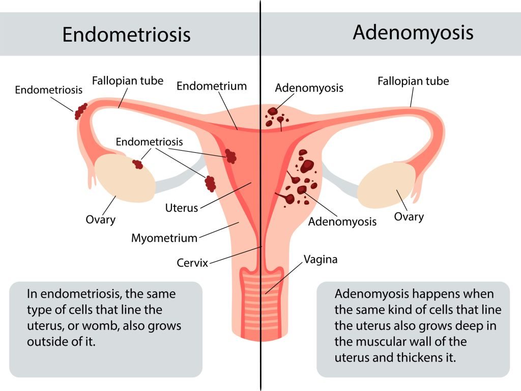
Adenomyosis happens when the tissue that lines your uterus grows into the muscle wall of the uterus itself. Every month that misplaced tissue does what uterine lining does — it swells and bleeds — but inside the muscle, with nowhere to go. The uterus becomes enlarged, tender and inflamed.
That mechanism explains the symptom pattern precisely: crushing cramps, heavy bleeding, and a deep dragging pain that hormonal treatments often can’t fully quiet. It also explains the detail that most distinguishes adenomyosis from ordinary period pain — cramps that have got worse year over year.
It is not endometriosis, though the two are constantly confused. In endometriosis, lining-like tissue grows outside the uterus. In adenomyosis it grows into the uterine muscle. About one in five women have both, and they need different treatment.
Three patterns — and the pattern decides your options
Adenomyosis doesn’t look the same in every uterus, and the difference matters more than most women are told.
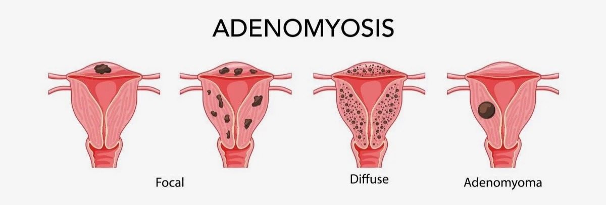
Hold onto those three words. When you get to the treatment list further down, which pattern you have rules some options in and others out — and it is the first question worth asking anyone who recommends something.
Fibroids and adenomyosis: nearly half of women have both
This is the part that quietly explains a lot of confusing appointments.
About 48% of women with adenomyosis also have fibroids. Not instead of. As well as. And because the two cause overlapping symptoms — heavy bleeding, pressure, pain — and both make a uterus look bulky, they are constantly mistaken for each other.
They are genuinely different things. A fibroid is a discrete, self-contained muscle tumour with a clear border and its own blood supply; you can point at it on a scan. Adenomyosis is lining tissue infiltrating the muscle wall, often with no clear border at all. An adenomyoma sits awkwardly between the two — adenomyosis behaving like a fibroid, and routinely reported as one.

Two practical consequences:
- A scan that found fibroids hasn’t ruled out adenomyosis. If you were told you had fibroids, had them treated, and the pain and heavy periods didn’t fully settle — that is a common story, and adenomyosis is a common reason for it.
- When both are present, they can be treated together. Embolization reaches both in the same session, because both depend on the same blood supply. Results for adenomyosis are strongest when the fibroids alongside it are treated at the same time.
Which is why the imaging question below isn’t box-ticking. Knowing exactly what you have — and how much of each — is the treatment plan. We compared every fibroid treatment in a separate article.
Why it took so long for anyone to say the word
For decades, the only way to diagnose adenomyosis was to examine a uterus after it had been removed. The diagnosis was made by a pathologist, after the fact. That single historical accident shaped everything: the prevalence figures, the awareness, and the odds that anybody looked for it in you.
You’ll find wildly different numbers quoted for how common it is — anywhere from 1% to 70% — because studies count different women with different tools. The largest analysis to date pooled nearly 200 million women across 127 studies. The line that matters if you’re reading this:
Among women being investigated for pelvic pain or heavy bleeding, 41–49% turn out to have adenomyosis.
Nearly half. And about 48% of women with adenomyosis also have fibroids, which is a large part of why the two get mistaken for each other on imaging.
And the reason your ultrasound was “normal”
Transvaginal ultrasound is almost always the first test you’ll be given — painless, no radiation, widely available. Pooled across studies it finds adenomyosis in roughly seven out of ten women who actually have it. Which means around one in four is missed.
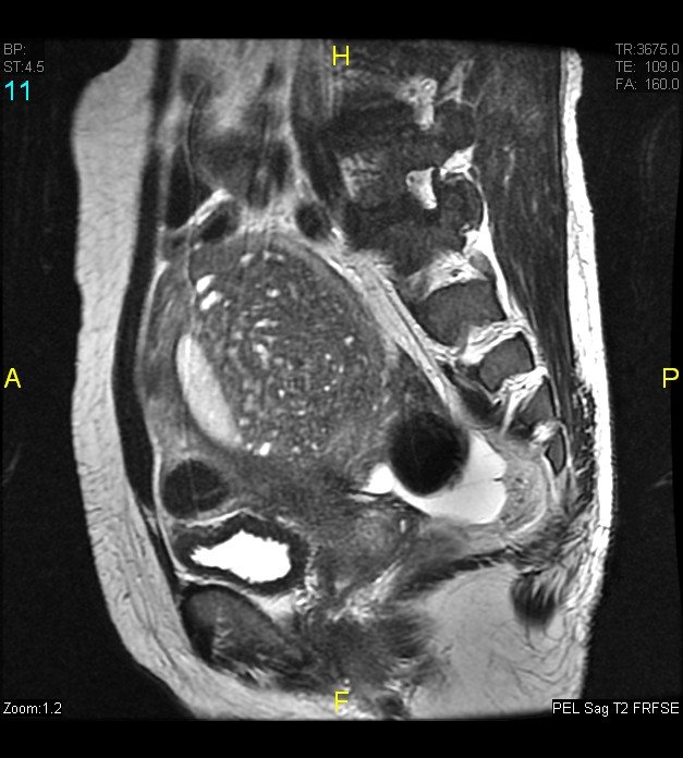
That isn’t a rare failure. It’s a known limit of the test. If you were told your ultrasound was normal and your symptoms haven’t changed, that is a reason to ask for an MRI — not a reason to accept that nothing is wrong.
MRI is the more reliable test. It measures the junctional zone — the border between lining and muscle, often the earliest sign there is — and it separates adenomyosis from fibroids, which matters when nearly half of women have both.
One thing that is not on the list of causes
Nobody fully knows why adenomyosis happens. What’s well established is that it runs on estrogen (symptoms track your cycle and usually settle after menopause), that previous uterine surgery may disturb the border between lining and muscle, and that it’s most often diagnosed in the 40s — though better imaging is now finding it in far younger women.
Note what is not on that list: anything you did. Adenomyosis is not caused by your diet, your weight, your stress, or waiting to have children.
Your real options — all of them
Hormonal treatment
The pill, the hormonal IUD, injections. These genuinely reduce bleeding and pain for some women, and they are a reasonable first step. Be clear about what they do, though: they mask the symptoms. They don’t treat the tissue causing them — and for many women with adenomyosis, they simply aren’t enough.
Hysterectomy
Removing the uterus removes the disease. It is definitive, and for some women it is the right choice — we say that plainly, and we refer for it. It is also major surgery, weeks of recovery, and final.
Surgical removal — but only for focal disease
When adenomyosis is concentrated in one spot rather than spread through the wall, it can form a distinct mass called an adenomyoma. Because it sits in one place, a surgeon can sometimes cut it out and repair the uterus.
The limits matter: it’s an option for focal disease only, not for the diffuse pattern, which is the more common one. It doesn’t treat adenomyosis elsewhere in the wall. And it is still surgery.
Uterine artery embolization
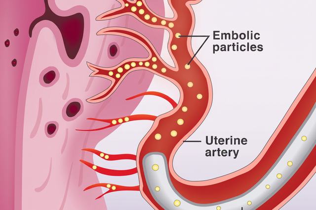
The third option, and the one most women are never told about. Through a pinhole in the wrist or groin, an interventional radiologist blocks the small arteries feeding the diseased tissue in the uterine wall. Starved of blood, that tissue shrinks and scars down — while your uterus stays where it is.
It is the only non-surgical treatment that addresses the tissue itself rather than masking the symptoms. It treats diffuse disease, focal adenomyomas, and the fibroids that nearly half of women have alongside it — in the same session.

What the research actually shows
Adenomyosis research is younger and thinner than the fibroid literature — there is still no large randomized trial — and we would rather show you the actual shelf than imply it’s taller than it is.
| Study | What it found |
|---|---|
| Australian durability study — the longest follow-up published | Of 91 women followed a mean of 52 months (and out to nearly ten years in some cases), 82 were still happy or very happy with the result, with no return of symptoms and no further procedure. Symptom scores fell from 58.9 to 20.0; quality-of-life scores more than doubled. |
| The largest pooled analysis — 1,049 women | 83% improved after embolization. |
| A five-year series of 252 women | Period pain still improved in 70%, heavy bleeding in 69% at five years. Durable for most — not for everyone. |
| QUESTA (2025) — head-to-head with hysterectomy | 101 women who chose either embolization or hysterectomy. Quality of life improved significantly after both, with comparable one-year scores — and 44 of the 50 women who chose embolization kept their uterus. |
One fear worth putting to rest: in the durability study, embolization did not appear to bring menopause forward.
And one thing we won’t claim: that this is a cure for adenomyosis. It isn’t. It is the only reliable uterus-sparing treatment for it. How long the improvement lasts generally depends on your age at treatment — adenomyosis runs on estrogen, so the closer you are to menopause, the lower the risk of recurrence. If symptoms do return, re-treatment can be considered.
Four questions to insist on before anyone recommends a treatment
- Is it diffuse or focal? It changes which options are even possible.
- Do I also have fibroids? Nearly half of women do — and it changes the plan, usually in your favor.
- Do I also have endometriosis? About one in five do. Treating one won’t fix the other.
- Am I anemic? Not “is my hemoglobin normal.” Ask for a ferritin.
And a principle underneath all four: being offered a hysterectomy for a symptom is not a plan. If heavy bleeding or pain is what sent you to the doctor and nobody has imaged you properly, then nobody yet knows what is causing it — and removing an entire organ to treat a symptom whose cause was never identified isn’t a treatment plan.
If this is the first time you’ve read your own symptoms described
That happens a lot on this page, and it is worth acting on. Bring your imaging, or ask us and we’ll order the MRI. What we’ll tell you at the end is which of these options fits what the scan actually shows — including the ones we don’t perform.
Read more about adenomyosis and embolization, or book a consultation.
Written and reviewed by the physicians of Arthritis Relief & Vascular Centers. This article is general health education, not medical advice, and reading it does not create a doctor–patient relationship. Every case is different — talk with a physician who has seen your imaging and examined you before making a treatment decision. If you are having a medical emergency, call 911.