

If a doctor has mentioned genicular artery embolization to you — or you found it while searching for anything that isn’t a knee replacement — the explanations you’ll find online tend to be either three sentences long or written for radiologists.
Here is the middle version: what it treats, what actually happens on the day, what the research shows, and who it isn’t for.
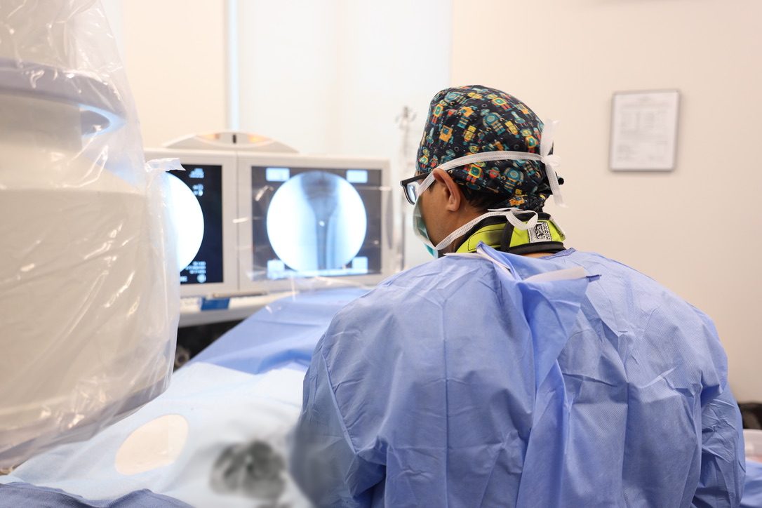
Start with what’s generating the pain
The standard story about knee arthritis is mechanical: cartilage wears out, bone rubs on bone, it hurts. That story explains some of the pain. It doesn’t explain the swelling, the throbbing at night when you aren’t standing on it, the good weeks and bad weeks, or why knees that look terrible on X-ray often feel fine.
What explains those is the joint’s lining — the synovium. In an arthritic knee it becomes chronically inflamed, and inflamed tissue grows a web of tiny abnormal blood vessels to sustain itself. New pain nerve endings grow alongside those vessels. The result is a patch of tissue inside your knee that is permanently irritated, permanently over-supplied with blood, and permanently wired to complain.
Embolization doesn’t treat the cartilage. It treats the blood supply keeping the inflammation alive.
So what is embolization?
Embolization is a technique interventional radiologists have used for decades: reach a target through the arteries with a catheter, then block the specific vessels feeding a problem. It is how fibroids are treated without a hysterectomy and how bleeding is stopped without an operation. Applied to the knee, the target is the abnormal vessel network feeding the inflamed lining.
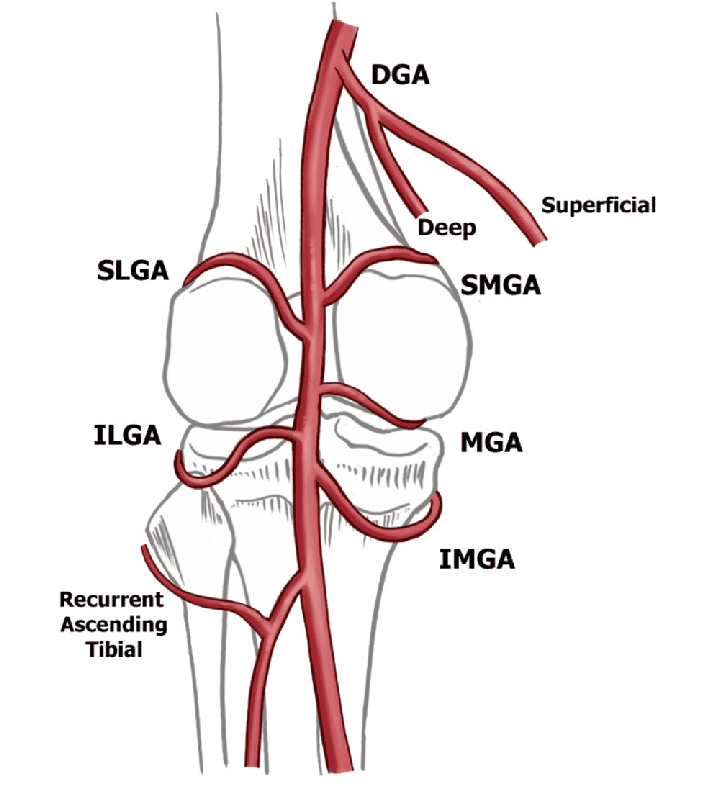
Genicular simply means “of the knee” — the genicular arteries are the small arteries that ring the joint. There are roughly eight of them, and in an arthritic knee some are feeding tissue that shouldn’t be getting that much blood.
What actually happens on the day
This is the part patients most often picture wrong. When people hear “catheter” and “artery” they imagine an operating room and a hospital stay. It isn’t that.
- You get comfortable. An IV delivers sedation — anti-anxiety medicine plus pain medicine. You’re relaxed and drowsy and breathing on your own. This is not general anesthesia.
- The pinhole. The doctor numbs a spot on the upper thigh and makes a pinhole opening in the artery. No incision, no stitches. The only thing most patients feel all day is that numbing pinch.
- The map. A catheter thinner than a phone-charger cord travels to the knee. Dye flows, and live X-ray (fluoroscopy) shows which vessels are abnormal — the dark “blush” of an inflamed lining is unmistakable once you’ve seen it.
- The treatment. Only the abnormal vessels are blocked. Some patients feel an ache like their own arthritis while it happens — which is oddly reassuring, because it usually means the right vessels are being treated. About 45 minutes on the table.
- Sealed and resting. The catheter comes out, the pinhole is sealed, and you rest 30 to 45 minutes purely for observation.
- Home. Your driver takes you. Back to normal daily activity right away, to tolerance; nothing heavy for a couple of days. Plan on three to four hours with us all told.
“Are you cutting off the blood supply to my knee?”
No — and it’s the question almost every patient asks, so it deserves a direct answer. The healthy arteries that nourish bone and normal tissue are left alone. Only the abnormal vessels that grew in response to years of inflammation are targeted, and the knee’s circulation has plenty of redundancy by design.
What gets injected, and does it stay there?
The blocking agent we use is a slurry of a medication (imipenem/cilastatin) mixed with X-ray dye. Together they form microcrystals smaller than a grain of sand, which lodge in the abnormal vessels — then dissolve into the bloodstream within 30 to 45 minutes. Nothing permanent stays in your body. In the study that compared these temporary crystals head-to-head against permanent beads, relief at two years was just as good.
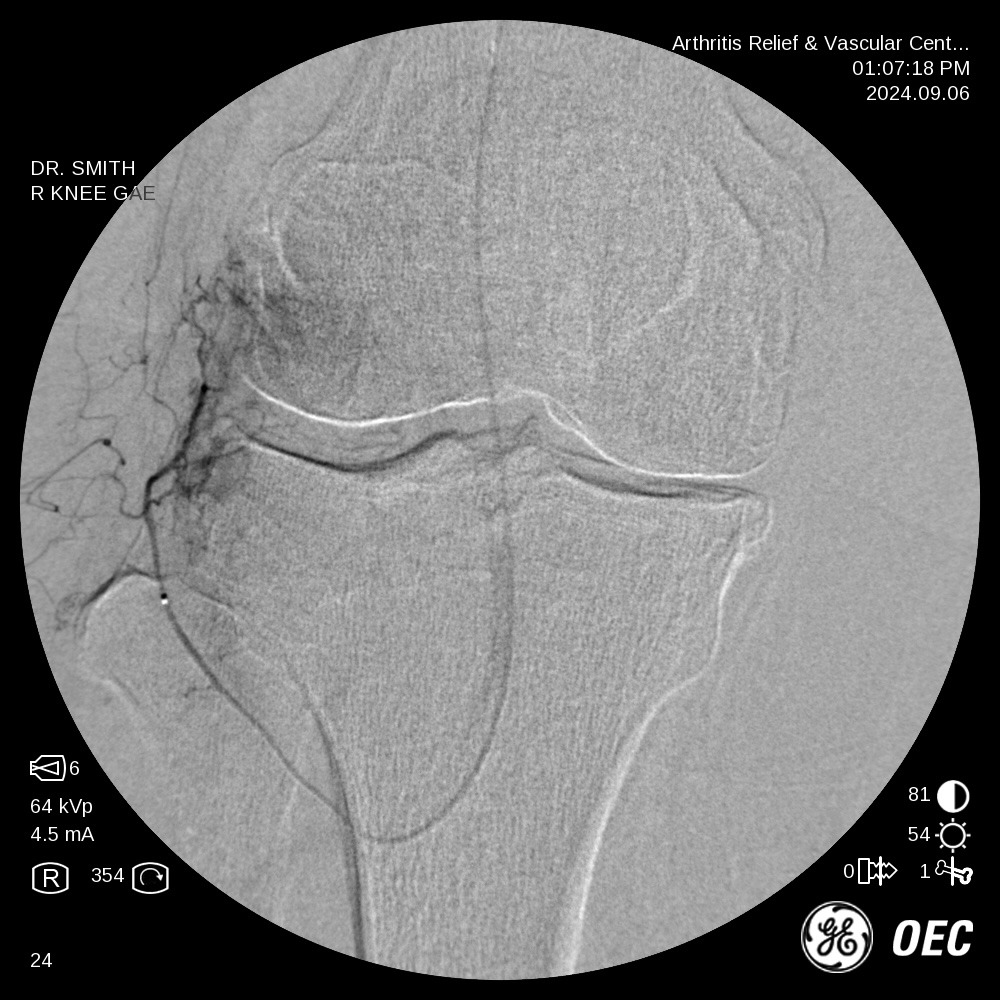
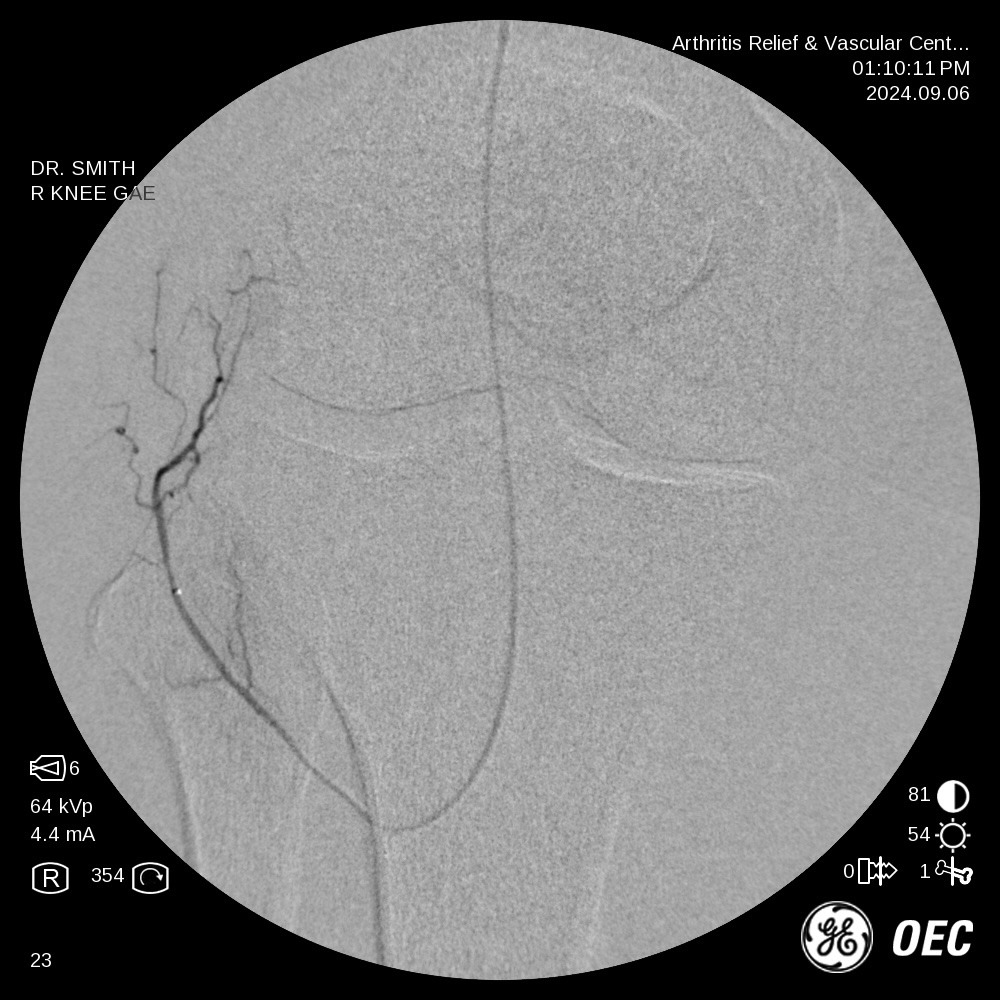
Angiograms from genicular artery embolizations performed at ARV Centers, San Antonio. Patient identifying information removed. Individual results vary.
What the research shows
This is where a lot of clinics get vague. We’d rather give you the numbers and their limits.
| Question | What the studies found |
|---|---|
| Is it just a placebo? | In the only US randomized sham-controlled trial, not one patient in the fake-procedure group improved — and every one of them chose to cross over and have the real treatment. About four in five of the treated patients improved within a month. |
| How well does it work? | Across nine clinical studies covering 270 patients, about three out of four had meaningful pain relief one year after the procedure. In a UCLA trial that deliberately included many severe, bone-on-bone knees, two-thirds cut their pain and symptom scores at least in half at one year. |
| How long does it last? | Among patients who responded, about seven in ten were still doing well at two years. Some symptoms can return between years one and two, and some patients choose a repeat treatment. |
| Does it lead to surgery anyway? | In the pooled studies, only about 1 patient in 20 went on to a knee replacement within two years. |
| Is it safe? | Across those 270 study patients, side effects were minor and short-lived — the most common was temporary skin discoloration near the knee, in about one patient in nine. Serious events were rare. |
The testing hasn’t stopped, either. The largest sham-controlled trial of GAE ever run — GENESIS 2, 110 patients randomized one-to-one, half receiving a placebo procedure — is underway right now. We’ll publish its results here when they come out, whatever they show. That is how a real treatment behaves.
What it doesn’t do
GAE does not regrow cartilage and it does not cure arthritis. It treats the inflammation that drives much of the pain. About one in five patients won’t respond meaningfully — and if we think your knee makes you a poor candidate, we will tell you before you spend a dollar.
It also closes no doors. In the two-year British trial, every patient who later chose a knee replacement had a normal operation with no added difficulty. Injections, a repeat embolization, and surgery all remain available afterwards.
Who it’s for
GAE may be right for you if you have knee arthritis pain that has lasted six months or more; injections, therapy or medication haven’t given lasting relief; and you’re not ready for a replacement — whether because you’re considered too young, because surgery is too risky for you, or because you simply don’t want one yet.
It isn’t right if your pain isn’t coming from arthritis, if you have certain circulation problems or advanced kidney disease, or if your knee genuinely needs an operation. We turn away patients who aren’t candidates for what we do, routinely.
Already had a knee replacement, and it still hurts?
That’s a different situation with a different workup — the implant has to be cleared by your surgeon first — and it’s become one of the most common reasons patients come to us. We wrote a full article on it.
The practical questions
Is it covered? By most insurance, including Medicare. We run a complimentary benefits check when you come in, so you know the number before anything is scheduled.
Do I need a referral? Not for your initial consultation.
Who performs it? A vascular and interventional radiologist — a physician trained to work inside blood vessels under image guidance. It’s worth asking any doctor how many they have personally done. Ours is more than 700 genicular artery embolizations.
If you want to know whether your knee is a candidate, bring your imaging and your questions. Read the full procedure page, or book a consultation.
Written and reviewed by the physicians of Arthritis Relief & Vascular Centers. This article is general health education, not medical advice, and reading it does not create a doctor–patient relationship. Every case is different — talk with a physician who has seen your imaging and examined you before making a treatment decision. If you are having a medical emergency, call 911.